Janet Hetherington talks to the innovators of the digital breast tomosynthesis (DBT), a futuristic mammogram procedure that may soon become the norm.

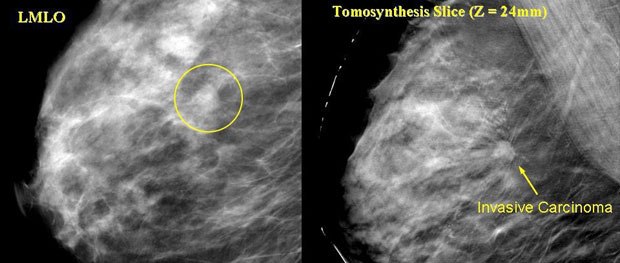
Technology has caught up with theory with mammograms. On the left is a standard 2D left medio-lateral oblique (LMLO) view with an obscure lesion while a tomosynthesis slice clearly shows a patent lesion. Courtesy of Mercury Computer Systems.
Back in the 1970s, Dr. Daniel Kopans, professor of radiology at Harvard University, first got the notion that digital tomosynthesis -- a 3D visualization technique that involves synthesizing multiple image slices -- could be used to study and diagnose breast cancer. However, Dr. Kopans had to wait for the technology to catch up with the theory -- and now, that day has arrived.
Dr. Kopans is currently overseeing a large screening study of some 3,000 women at Massachusetts General Hospital, using a prototype digital breast tomosynthesis (DBT) imager (a 3D mammogram procedure). Basically, DBT constructs a 3D map of the breast to help radiologists see tumors that might be obscured on 2D scans. While traditional mammography uses two X-ray views per breast, DBT integrates up to 25 views per breast, each taken from a different vantage point along an arc.
Massachusetts General Hospital holds the DBT patent, which it licensed to General Electric (GE) so a working prototype could be built.
"A standard mammogram examination captures a single image of an entire breast," says Dr. Kopans. "The problem is that everything in the breast, from one side to the other, is superimposed into one 2D view. DBT enables us to look into a 3D visualization of the breast. Individual layers in the breast tissue are visible separately from overlying and underlying structures, making it harder for cancers to hide."
Mammography normally requires two X-rays of each breast taken from different angles -- top to bottom and side to side. The breast is pulled away from the body, compressed and held between two glass plates to ensure that the entire breast is viewed. Regular mammography records the pictures on film, while digital mammography records the pictures on a computer. A radiologist reads the images. Breast cancer, which is denser than most healthy breast tissue, appears as irregular white areas, which are sometimes called "shadows."
DBT allows multiple X-ray pictures of each breast to be taken from many angles. The breast is positioned the same way it is in a conventional mammogram. The X-ray tube moves in an arc around the breast while multiple images are taken during the examination. Then the information is sent to a computer, where it is assembled to produce a 3D representation.
"Current film-based or 2D digital mammography takes an image of a 3D object (the breast) and projects it into 2D," comments Colin Murphy, director of sales and marketing for Mercury Computer Systems, a firm that is working closely with Massachusetts General Hospital on the project. "3D positioning of structures within the breast is lost and structures along the path of the rays from the emitter to the detector occlude each other. DBT digitally reconstructs a 3D volumetric representation of the breast, so it can be viewed in 3D, with all the spatial relationships intact and without the inherent 2D occlusion problem."
Dr. Kopans further explains that since breast tissue can be dense, being able to view the image in 3D makes it easier to detect both cancerous tumors and benign growths. The Massachusetts General Hospital technique eliminates superimposed normal breast tissue that can obscure cancers on standard mammograms. This allows for improved detection and earlier treatment in the tumor growth cycle. "The beauty of this technology is that all the regular rules of a mammogram apply, but you can see things better," Dr. Kopans adds.

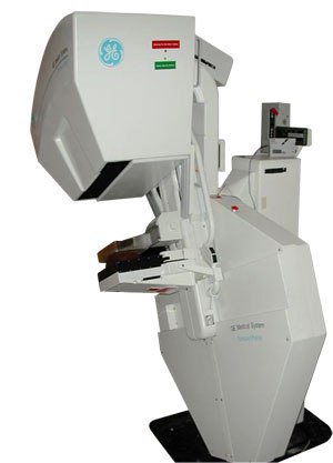
Made possible by a grant from the U.S. Department of Defense and the engineering expertise of the General Electric Co., a tomosynthesis scanner was built and installed at the Massachusetts General Hospital. Courtesy of Mercury Computer Systems.
Humble Beginnings
The DBT imager prototype being used in the trial is actually the second one to be built. The first one was fashioned by GE, using grant money from the U.S. Department of Defense, which provides funding for breast cancer research.
"It took all night to process one file," Dr. Kopans says. "It was not clinically practical." For the second imager prototype, also built by GE, a 32-processor cluster system was implemented, and results improved. "Parallelizing the problem meant a reduction of the process to 15 minutes," says Dr. Kopans.
Then, the Massachusetts research team turned to Mercury Computer Systems. "Mercury has a long history -- 25+ years -- of solving difficult computing problems in medical, defense and commercial applications," comments Mercury's Murphy. "Massachusetts General Hospital approached us because of our history of success, both with them on previous projects, and with other medical OEMs. The Hospital knew Mercury had a broad portfolio of solutions (based on PowerPCs, FPGAs, DSPs and GPUs) and could develop a suitable solution that met their criteria -- performance, cost and size."
While Mercury Computer Systems came to the project with a good appreciation of the reconstruction technology necessary for low-contrast imaging data, the company had not worked directly on a digital mammography project before. "The goals were to provide 3D reconstructed data that was diagnostically equivalent or superior to the data produced by Massachusetts General Hospital's Pentium code, but with vastly improved reconstruction time and a corresponding decrease in hardware costs," notes Murphy.
Murphy says that systems consulting engineer Iain Goddard was the key individual at Mercury who closely interacted with the Hospital research staff. "He worked with their scientists to migrate the parallelized algorithm that Massachusetts General Hospital was utilizing onto the Mercury GPU platform," Murphy says. "Mr. Goddard was assisted by principal systems engineer Scott Thieret in the implementation aspects of the project."
Mercury's objective was to make the DBT processing practical. Instead of using a cluster of 32 PCs for the computationally intensive image-processing algorithm, Mercury designed a solution based on NVIDIA graphics technology -- a key element, providing near realtime results. Mercury Computer Systems initially implemented on the ATI 9800, the highest performance off-the-shelf GPU board available at the time.
"However, as Mercury's relationship with NVIDIA grew, Mercury moved to NVIDIA's professional product line," Murphy says. "These devices had more memory and a longer MTBF than the high-end gaming cards."
Murphy explains that while the algorithm is implemented in a way that will scale across several GPUs simultaneously with a linear speedup, the performance of the Quadro FX4500 proved sufficient to meet Massachusetts General Hospital's performance requirement.
The Need for Speed
The results are impressive. A single NVIDIA Quadro graphics board proved able to generate imagery as fast as the Massachusetts General Hospital's 32-processor cluster -- which occupied half a room -- in under five minutes. That's more than 60 times faster than the previous single-system solution.
"In addition to being more compact in physical size, the Quadro FX4500 pulls much less power from wall outlets and there's much less heat generated," notes Sanford Russell, professional market development manager at NVIDIA.

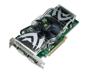
The Quadro FX 4500 generates imagery as fast as the Massachusetts General Hospital's 32-processor cluster -- which occupied half a room -- in under five minutes, 60 times faster than the previous single-system solution. © NVIDIA.
Since that time, NVIDIA Quadro graphics and PCI Express technology have enabled the latest solution to reduce the image processing time another 50%.
"There's a seemingly unquenchable thirst for processing power," Russell says. "There's a tremendous amount of data to process. There's stacking of multiple slices, with math required to fill out the gaps. The result is that you can turn the image around and explore it, looking behind dense tissue to find cancer."
"The speed of image acquisition is very important in capturing the tomographic data used for the reconstruction of the breast tomosynthesis data," comments Mercury's Murphy. That's because rapid computation, combined with compact sized and affordable equipment, makes the widespread implementation of DBT technology more viable.
"Dr. Kopan's vision is that once a DBT is taken, before a patient leaves, the doctor will have the results immediately and be able to consult with that patient," NVIDIA's Russell offers.
Dr. Kopan reports that preliminary data from his study suggests that the DBT procedure may also reduce "false positive" findings.
Comfortable Fit
DBT may provide another benefit -- more comfort. "From a woman's perspective, there's nothing really different from a regular mammogram," Dr. Kopans says. "The images are obtained the same way. However, this method requires that the breast be squeezed only once. The research suggests that women have found tomosynthesis to be more comfortable."
The procedure seems familiar to patients, and to the professionals who operate the equipment as well. When it comes to teaching radiologists how to use DBT, Dr. Kopans says, "The learning curve is very short."
"The system has been utilized by the experienced mammographers at Massachusetts General Hospital and it has been well accepted by them in their clinical use," comments Mercury's Murphy. "It is likely that one of the goals of the clinical validation undertaken by the OEM utilizing the system is to obtain user feedback on its usability for radiologists and RTs."
As for saving the captured images, the process is similar to other medical procedures. "Storing is the same way as for MRIs and CT scans," says NVIDIA's Russell. "It's the processing that's different."
A Caring Technology
Russell makes a point of saying that for all involved, the DBT work is very important on a personal level. "Dr. Kopans is really helping people," he says.
Almost a quarter of a million women in the U.S. alone are diagnosed with breast cancer every year. Early detection is a vital strategy and the major proven method for reducing breast cancer mortality.
While the DBT is still in clinical trials, Dr. Kopans says the next step is to gain FDA approval, which is anticipated within the next year. He also notes that while Massachusetts General Hospital holds the DBT patent, other facilities are in "research mode" to develop competing technology. "Other people are working around the patent," he says. The technique has also been written up in a number of journals.
Once FDA approval is obtained, DBT imaging may soon become the norm. As NVIDIA's Russell observes, "All of the medical industry is moving to 3D visualization."
Janet Hetherington is a freelance writer and cartoonist who shares a studio in Ottawa, Canada, with artist Ronn Sutton and a ginger cat, Heidi.


